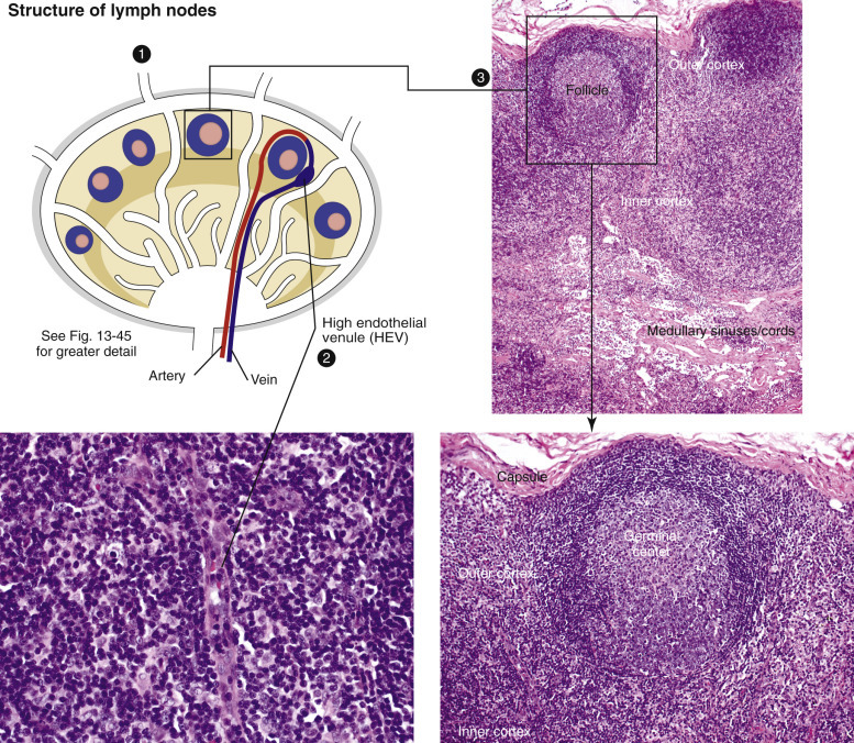
Figure 13-46.

Cellular Zones of a Lymph Node.
1, Antigen arrives in the afferent lymphatic vessels, empties into the subcapsular sinus, and drains into the trabecular and medullary sinuses. 2, As antigens travel through the sinuses, they are captured and processed by macrophages and dendritic cells (DCs), or antigen-bearing DCs in blood can enter through high endothelial venules (HEVs). B lymphocytes encounter DCs charged with antigen, are activated, and migrate to a primary follicle to initiate germinal center formation, creating secondary follicles. 3 and lower right image, lymphoid follicles. Germinal centers have a distinct polarity (superficial or light zone and a deep dark zone), and the mantle cell rim partially encircles the germinal center and is wider over the light pole of the follicle.
(Courtesy Dr. A.C. Durham, School of Veterinary Medicine, University of Pennsylvania; Dr. M.D. McGavin, College of Veterinary Medicine, University of Tennessee; and Dr. J.F. Zachary, College of Veterinary Medicine, University of Illinois.)
