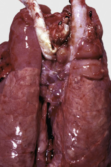
Figure 13-75.

Acute Lymphadenitis, Tracheobronchial Lymph Nodes, Pig.
The tracheobronchial lymph nodes are draining the cranial lung lobes, which are consolidated due to severe pneumonia. The nodes are enlarged and reddened. This appearance is due to the “reversed” anatomic arrangement in the pig lymph node; the blood-filled sinuses are obvious at the surface.
(Courtesy Dr. M.D. McGavin, College of Veterinary Medicine, University of Tennessee.)
