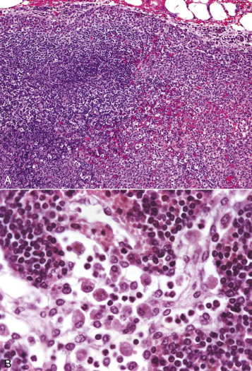
Figure 13-78.

Acute Lymphadenitis (Early), Lymph Node, Dog.
A, The sinuses and the parenchyma of the cortex and medulla have coalescing foci of neutrophilic inflammation, necrosis, hemorrhage, and fibrin deposition. H&E stain. B, The medullary sinus contains numerous macrophages (sinus histiocytosis) and fewer neutrophils. This is the type of early response seen when a lymph node drains an inflamed area. Medullary cords are filled with lymphocytes and plasma cells. H&E stain.
(A courtesy Dr. A.C. Durham, School of Veterinary Medicine, University of Pennsylvania. B courtesy Dr. H.B. Gelberg, College of Veterinary Medicine, Oregon State University.)
