
Figure 13-91.
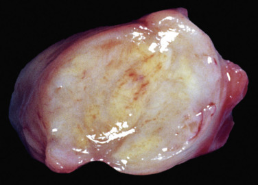
Mesenteric Lymph Node, Diffuse Granulomatous Lymphadenitis, Histoplasmosis, Dog.
The lymph node is enlarged, the cut surface shows loss of architecture, and the tissue bulges because of the diffuse granulomatous inflammation (see Fig. 13-92).
(Courtesy Dr. M.D. McGavin, College of Veterinary Medicine, University of Tennessee.)