
Figure 2-25.
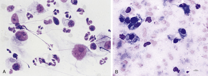
A, Reactive fibroplasia. Tissue scraping. Cat. Oral mass with associated septic inflammation. Pictured are several plump mesenchymal cells with a stellate to spindle appearance and prominent nucleoli along with suppurative inflammation. The severity of the inflammatory response warrants caution in suggesting a malignant mesenchymal mass or sarcoma. Note the nuclear streaming appears as purple strands. (Aqueous Romanowsky; HP oil.) B, Postnecrosis fibroplasia. Tissue aspirate. Dog. Facial swelling related to myositis. The background contains amorphous grey material supportive of necrosis. Several fibroblasts indicate a reparative process following tissue damage. (Modified Wright; HP oil.)