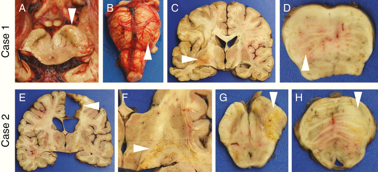
Fig. 1.
Gross findings in postmortem GBMs. At the time of brain removal in Case 1, separation of the posterior fossa contents via midbrain transection revealed a swollen, discolored left cerebral peduncle (a, arrowhead) and left pons (b, arrowhead). After formalin fixation, coronal sections showed a relatively small area of hemorrhage and necrosis in the left temporal lobe (c, arrowhead), but no notable mass effect or midline shift. Axial sections of the brainstem again found the left pons to be swollen and discolored (d, arrowhead). In Case 2, the original tumor site in the right frontal lobe showed a resection cavity surrounded by glial scar (e, arrowhead), as well as streaks of yellow necrosis through the internal capsule headed toward the right cerebral peduncle (f, arrowhead). No mass effect was noted. Sections of the midbrain (g) and pons (h) revealed swelling and necrosis in the right cerebral peduncle and basis pontis, respectively (arrowheads). In (d), (g), and (h), anterior is up.

