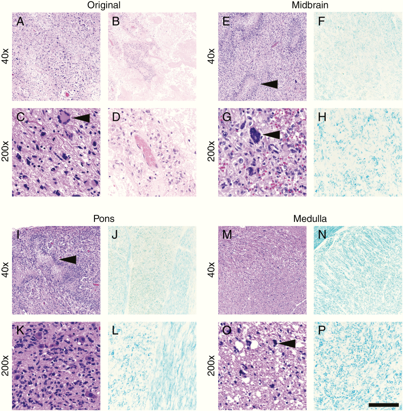
Fig. 2.
Case 1 histology. The original tumor site in the left temporal lobe showed large areas of viable tumor (a) and therapy-induced cellular atypia (c, arrowhead), as well as abundant therapy-related necrosis (b, d). In contrast, sections of the midbrain (e, g) and pons (i, k) showed viable tumor with pseudopalisading necrosis (e and i, arrowheads). At higher magnification, scattered tumor nuclei with therapy-associated atypia were observed (g, arrowhead). Tumor infiltration was heavy around pontine neurons (k). In the medulla, overall tumor burden was lighter (m, o), but still readily apparent (o, arrowhead). LFB highlighted massive loss of myelination in the midbrain (f, h), pons (j, l), and medulla (n, p). Compare with the undamaged white matter in Fig. 4. Scale bar in (p) = 400 µm at 40x and 80 µm at 200x.

