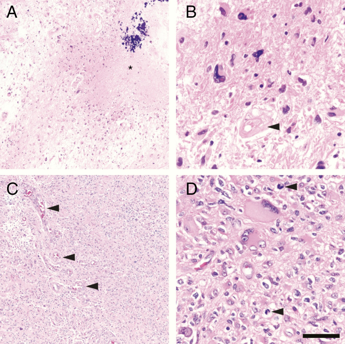
Fig. 1.
“Residual” versus “recurrent” GBM. (A) Patterns more suggestive of residual, posttherapy GBM include low-to-moderate cellularity tumor with large swaths of necrosis that may contain mineralization (*). (B) At higher power, the tumor cells still may be alive, but do not appear healthy. Such cells usually show marked nuclear atypia with very few mitoses, if any. Blood vessels often appear devitalized, hyalinized, and distorted (arrowhead). (C) Genuinely recurrent GBM, on the other hand, is more densely cellular, often with robust microvascular proliferation (arrowheads). (D) While recurrent GBM will still contain highly atypical cells, most cells will appear quite viable, with scattered mitoses (arrowheads). Scale bar = 250 µm in (A) and (C), and 50 µm in (B) and (D).

