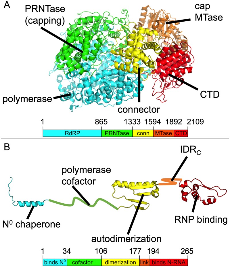
FIG 1.
Structures of the VSV L and P proteins. (A and B) VSV L protein (PDB ID 5A22) (A) and VSV P (a composite of PDB ID 3HHW, 2FQM, and 3PMK) (B) are shown in cartoon format with domains labeled and colored individually. Domain boundaries by residue number are provided below each cartoon. conn, connector. IDRc, C-terminal intrinsically disordered region.

