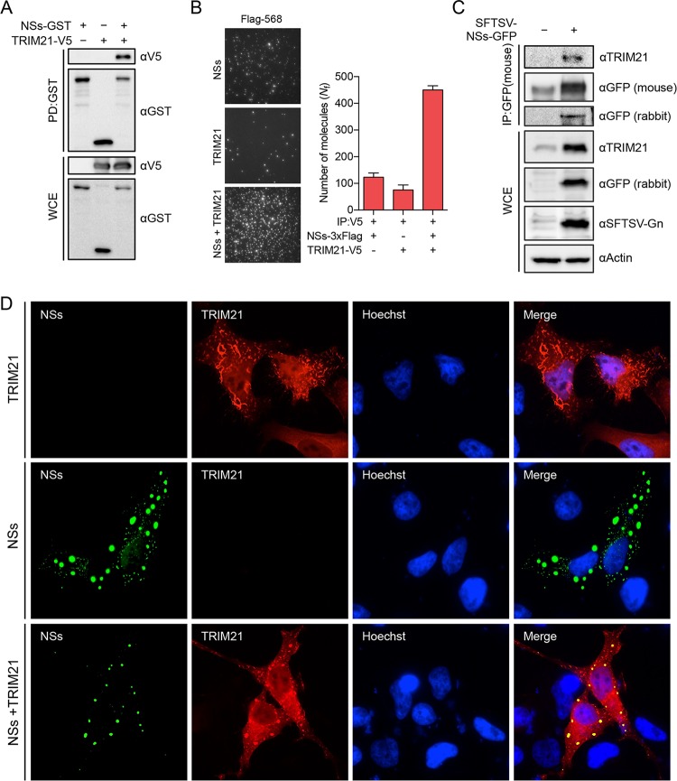
FIG 1.
NSs interacts with TRIM21. (A) HEK293T cells were transfected with NSs-GST and TRIM21-V5, and whole-cell extracts (WCEs) were pulled down by glutathione beads, followed by immunoblotting with the indicated antibody. (B) HEK293T cells were transfected with NSs-3×Flag and TRIM21-V5, and WCEs were applied to SiMPull analysis. (Left) Three representative images. (Right) Molecular numbers, in which the bar graphs indicate the average number of fluorophores per image. Error bars represent the SD of the mean across >20 images. The results of three independent experiments are represented. (C) RAW 264.7 cells were infected with SFTSV-NSs-GFP, a recombinant virus expressing GFP-tagged NSs, for 24 h and subjected to immunoprecipitation (IP) with anti-GFP antibody to pull down the GFP-NSs complex, followed by immunoblotting with anti-TRIM21 antibody to detect endogenous TRIM21. (D) HeLa cells were transfected with NSs-3×Flag-GFP and TRIM21-V5. The cells were fixed and stained with primary mouse anti-V5 antibody and with secondary Alexa Fluor 568-conjugated anti-mouse IgG antibody for confocal microscopy. Hoechst staining was used for the nucleus. The microscope images represent those from three independent experiments.

