
FIG 2.
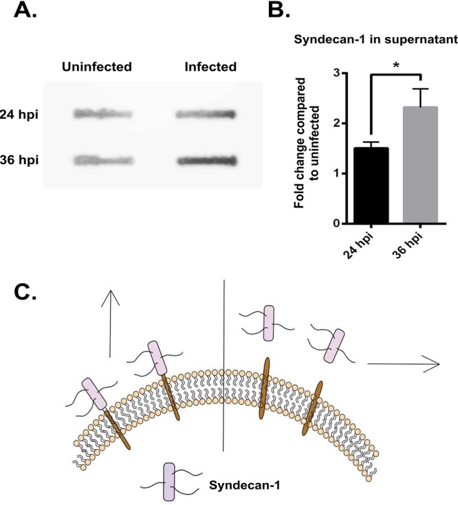
HSV-1 infection causes the shedding of syndecan-1 from the cell surface. (A) Slot blot assay shows increased syndecan-1 shedding in HCE cells. HCE cells were infected with KOS-WT at an MOI of 0.1 for 24 and 36 h. The supernatant was used to measure the levels of syndecan-1 that was shed from the cell surface. (B) Densitometry quantification of syndecan-1 shed in supernatant based on slot blot assay. The fold change was normalized to uninfected mock samples at each time point. (C) Image representing shedding of syndecan-1. The left side of the perpendicular line shows syndecan-1 bound to the cell surface, which was measured by flow cytometry. The right side of the perpendicular line shows syndecan-1 shed in the supernatant, which was measured by slot blot assay and is described above in panel A.