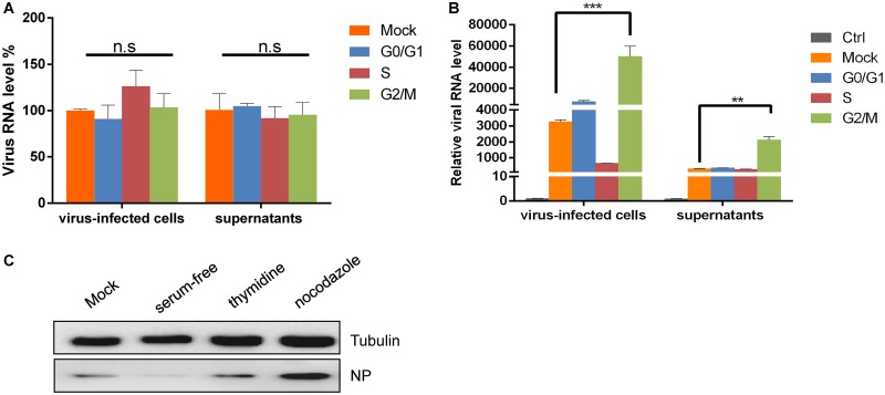
FIG 3.
G2/M phase synchronization promotes SFTSV proliferation. (A) HepG2 cells were cultured in complete medium, serum-free medium, medium containing 0.85 mM thymidine, or medium containing 50 ng/ml nocodazole for 24 h and then infected with SFTSV (0.1 MOI) for 30 min. The cells and supernatants were collected immediately and analyzed by qRT-PCR. (B) HepG2 cells were treated as described above. After infection with SFTSV (0.1 MOI), fresh complete medium, serum-free medium, medium containing 0.85 mM thymidine, or medium containing 50 ng/ml nocodazole was added to the culture and left for another 48 h. The cells and supernatants were collected for qRT-PCR analysis (n.s, P > 0.05; **, P < 0.01; ***, P < 0.001). (C) HepG2 cells were treated as described above but infected with SFTSV at an MOI of 1. After 48 h, Western blotting was performed to detect NP expression.

