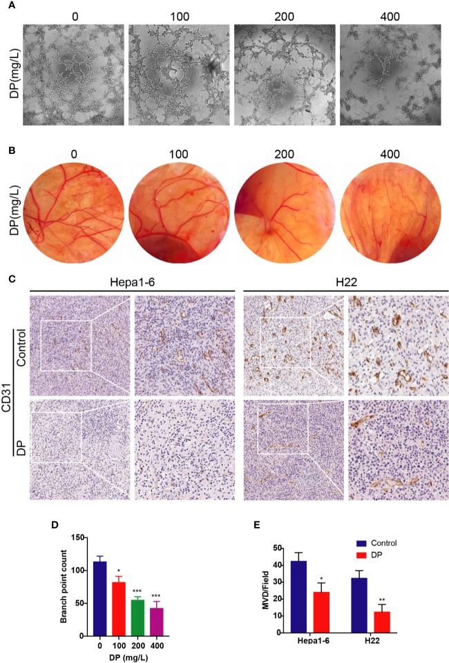
Figure 1.
DP inhibits angiogenesis in vivo and in vitro. (A, D) Tube formation of HUVECs on Matrigel. Cells were treated with CM derived from 0, 100, 200, and 400 mg/L DP treated HepG2 cells for 48 h. (B) The newly formed blood vessels on the chick embryo chorioallantoic membrane (CAM) after treated with DP (0, 100, 200, and 400 mg/L) for 48 h. (C, E) Histopathological analyses of tumor growth in mice xenografted with Hepa1-6 and H22 cancer cells. The tumor sections were subjected to IHC staining using an antibody against CD31. The magnification is 400x. Error bars represent mean ± SD from three independent experiments. (*P < 0.05, **P < 0.01, ***P < 0.001).

