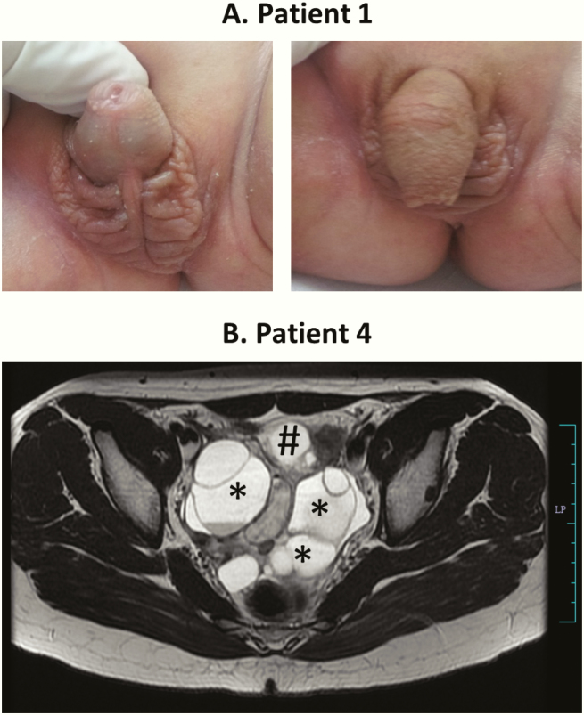
Figure 3.
Phenotype of girls with CYP19A1 mutations. A, Picture of the ambiguous genitalia at birth found in the 46,XX disorder of sex development index patient 1. Note the phallus-like tubercle with a single meatus opening at the base and the complete labioscrotal fusion (Prader IV). The photograph is shown with the permission of the parents. B, Magnetic resonance imaging of the pelvis in patient 4 at age 14 years showing a large, multiloculated, cystic mass (6.8 × 9.6 × 6.5 cm) involving bilateral adnexa (*), displacing the uterus (#) anteriorly and inferiorly. Few of the cysts are showing hemorrhage. Both ovaries cannot be separately visualized.

