
Figure 1.
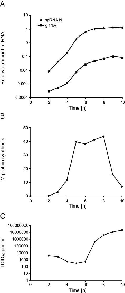
Analysis of the MHV infection progression by monitoring the viral RNA and protein synthesis, and the extracellular release of viral particles. HeLa‐CEACAM1a cells were infected with MHV‐A59 and the culture supernatants and cell lysates were collected at different p.i. time points as described in Experimental procedures. A. The total RNA was isolated from the infected cells and the amount of gRNA and sgRNA was quantified by RT‐PCR. Results are expressed using arbitrary units. B. Infected cells were metabolically labelled 30 min prior of being collected and lysed. Cell lysates were then immunoprecipitated with an antiserum again the complete MHV in combination with anti‐M protein antibodies. Finally, immuno‐complexes were resolved by SDS‐PAGE and the amount of radioactive M protein was quantified. Results are expressed using arbitrary units. C. The production of the progeny virus was assessed by determining the virus titre of the culture supernatants by end‐point dilutions on LR7 cells and then calculating the TCID50 units per ml of supernatant.