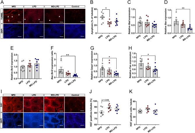
Figure 4.
Impact of NPD, LPD and MD-LPD on testicular apoptosis. Representative TUNEL and DAPI stained sections of NPD, LPD and MD-LPD testis and negative control (bar = 100 µm) (A) showing apoptotic cells (white arrow heads) and the percentage of tubules showing apoptotic cells (B). Relative testicular expression of BCL2-associated agonist of cell death (Bad) (C), BCL2-associated X protein (Bax) (D), B cell leukemia/lymphoma (Bcl2) (E), Bax:Bcl2 expression ratio (F), caspase 1 (Casp1) (G) and TNF receptor superfamily member 6 (Fas) (H). Representative Ki67 and DAPI stained sections of NPD, LPD and MD-LPD testis and negative control (bar = 100 µm) (I) with the percentage of tubules showing positive stained cells (J) and mean number of Ki67 positive cells per tubule (K). Data are mean ± s.e.m. n = 8 males per dietary group. Data were analysed by one-way ANOVA followed by Bonferroni post-hoc test, or Kruskal–Wallis test with Dunns multiple comparison test where appropriate. *P < 0.05, **P < 0.01.

 This work is licensed under a