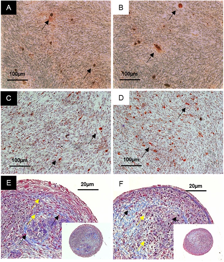
Fig 3. Tri-lineage differentiation of MSCs.
MSCs differentiated using hPL-PIPC are shown in A, C, and E, while MSCs differentiated using hPL-PIPL are shown in B, D, and F. A and B show Alizarin Red S staining, used to demonstrate mineralization (black arrows) after 28 days of stimulation in osteogenic medium. C and D show Oil Red O staining, used to demonstrate accumulation of lipid droplets (black arrows) after 14 days of stimulation in adipogenic medium. E and F show Masson’s trichrome staining, used to demonstrate collagen fibers (black arrows) and lacunae formation (yellow arrows) after 35 days of chondrogenic stimulation.

