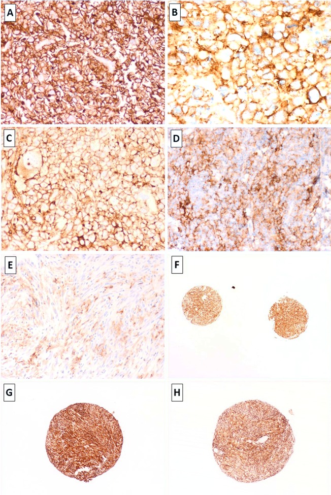
Fig 2. Strong membranous expression for PD-L1 idenified in leiomyosarcoma (LM-75), undifferentiated pleomorphic sarcomas (B,C: UPS-67 & UPS-11, respectively) and metastatic angiosarcoma (D, AS-2).
Focal heterogenous expression for PD-L1 in leiomyosarcoma (E, LM99). Concordant expression for PD-L1 in replicate cores was identified (e.g. 1F; UPS-21). Primary and recurrent UPS (UPS-61 and UPS-62: G & H) demonstrating concordant strong expression of PD-L1 in 100% of the tumour cells in both, the primary lesion and its recurence (TMA cores). Images taken at 2x – 40x magnification and stained with the PD-L1 SP263 clone, Ventana.

