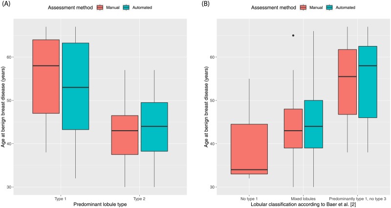
Fig 5. Boxplots demonstrating the association of qualitative terminal ductal lobular unit involution measures and age.
(A) Women with predominantly type 1 lobules were significantly older than women with predominantly type 2 lobules (manual method: p<0.01; automated method: p = 0.01). No woman presented with predominately type 3 lobules. (B) Women with “Predominantly type 1, no type 3” lobules were significantly older than women with “Mixed lobules” (manual method p<0.01; automated method p<0.01). No woman was assessed as having “No type 1” lobules by the automated method. The manual qualitative measures were obtained by consensus vote. The boxplots show the median value, interquartile range (IQR), and 5th and 95th whiskers.

