
Figure 11.
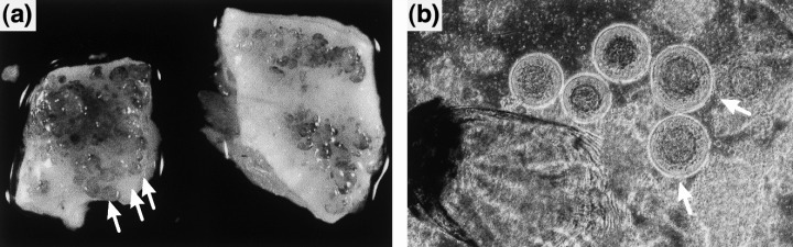
Lymphocystis disease virus (LCDV) infection in flounder. (a) Skin of LCDV‐infected flounder exhibiting multiple lymphocystis nodules (see arrows). (b) Explantats of LCDV‐infected flounder skin: lymphocystis cells are round and hypertrophied (see arrow), and 1000 times the size of uninfected cells (× 22). (The fish was kindly provided by Dr Steinhagen, Faculty of Veterinary Medicine, Hannover, Germany.)