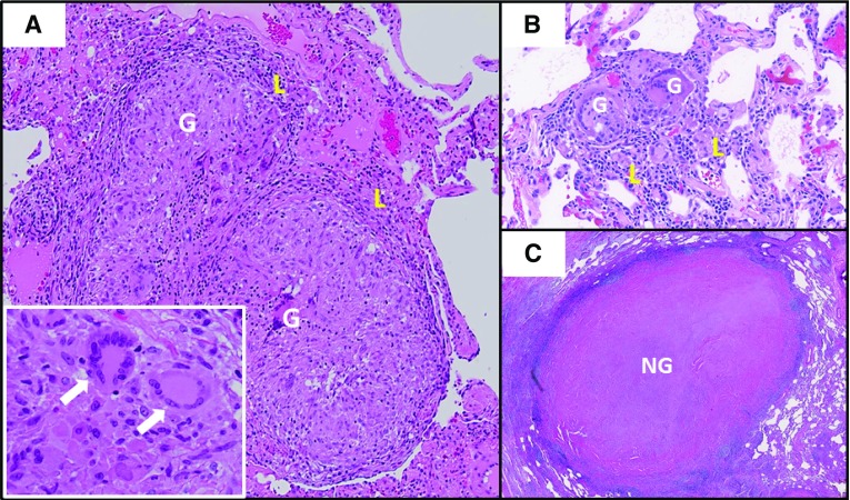
Figure 1.
Comparison of pulmonary sarcoidosis granuloma histology to other granulomatous lung diseases. (A) Typical sarcoidosis histology with well-formed granulomas comprised of macrophage aggregates (G) and featuring multinucleated giant cells (white arrows, inset), with minimal surrounding lymphocytic inflammation (L). (B) Hypersensitivity pneumonitis featuring smaller granulomas (G) with more extensive surrounding lymphocytic alveolitis (L). (C) A large acellular necrotizing granuloma (NG) caused by pulmonary Histoplasma capsulatum infection.

