
Figure 4.
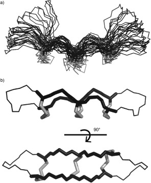
NMR structures of θ‐defensins. a) Ensemble of the 20 lowest‐energy structures of RTD‐1 (PDB 1HVZ)75 with disulfide bonds in grey. This was the first θ‐defensin structure published and suggested flexibility of the turn region. b) The symmetrical θ‐defensin BTD‐2 (PDB 2LYE)7 with the cyclic cystine ladder motif shown as sticks.