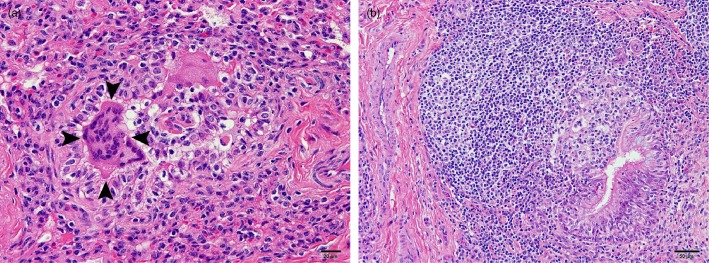
Figure 2.

(a) Sample of the lung of a steer infected with bovine coronavirus showing multinucleate epithelial syncytial cells in a collapsed bronchiole (arrowheads). (b) Sample of the lung of a calf infected with bovine coronavirus showing marked hyperplasia of the bronchiole‐associated lymphoid tissue, with hyperplasia and focal disruption of the bronchiolar epithelium by infiltrating lymphocytes. (Haematoxylin and eosin staining.)
