Figure 6.

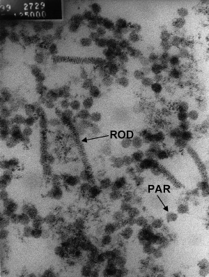
Transmission electron microscopy of vesicle showing particles (PAR) of 50–96 nm in diameter and rod‐shaped structures (ROD) 155–207 nm long (Smith 2000).

Transmission electron microscopy of vesicle showing particles (PAR) of 50–96 nm in diameter and rod‐shaped structures (ROD) 155–207 nm long (Smith 2000).