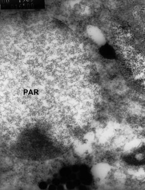
Figure 7.

Transmission electron microscopy of vesicles within nerve cells of the fasciculated zone of the eye of moribund Penaeus monodon. The vesicles are 3 μm in diameter and contain unidentified particles (PAR) 20 nm in diameter. Some particles also appear to be free in the cytoplasm (Smith 2000).
