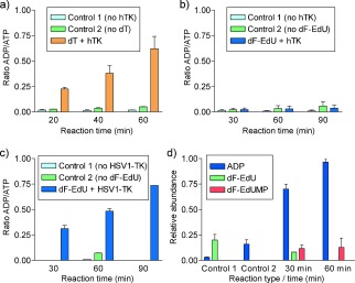
Figure 1.

a, b) In vitro phosphorylation of dT (a) or dF‐EdU (b) by hTK. c, d) Phosphorylation of dF‐EdU by HSV1‐TK according to the relative quantities of ADP, ATP, dF‐EdU, and dF‐EdUMP. In all panels, Control 1 lacks the kinase and Control 2 lacks the nucleoside.
