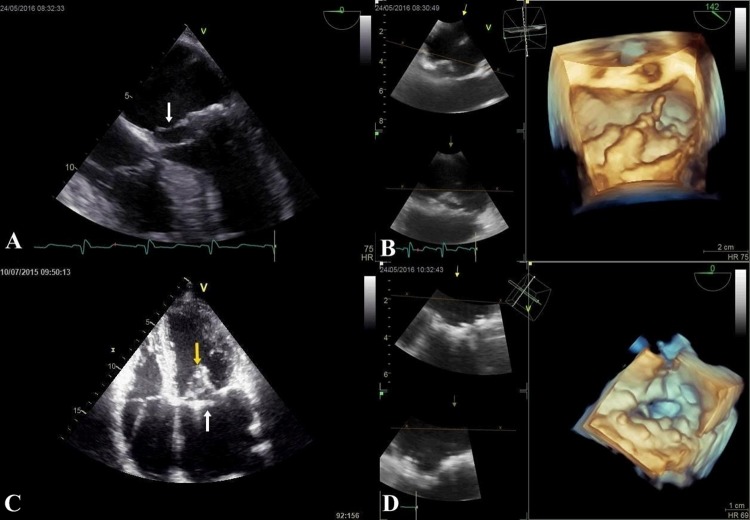
Figure 4. 2D and 3D assessment of patient with MVP.
A. 2DTEE pre-operative imaging showing flail posterior leaflet (arrow); B. 3DTEE pre-operative imaging showing flail posterior leaflet( P2); C. 2D TTE apical four chamber view - post-operative imaging showing perfect coaptation (yellow arrow) and lack of leaflet prolapse (white arrow); D. 3DTEE post-operative imaging showing perfect coaptation and lack of leaflet prolapse.

