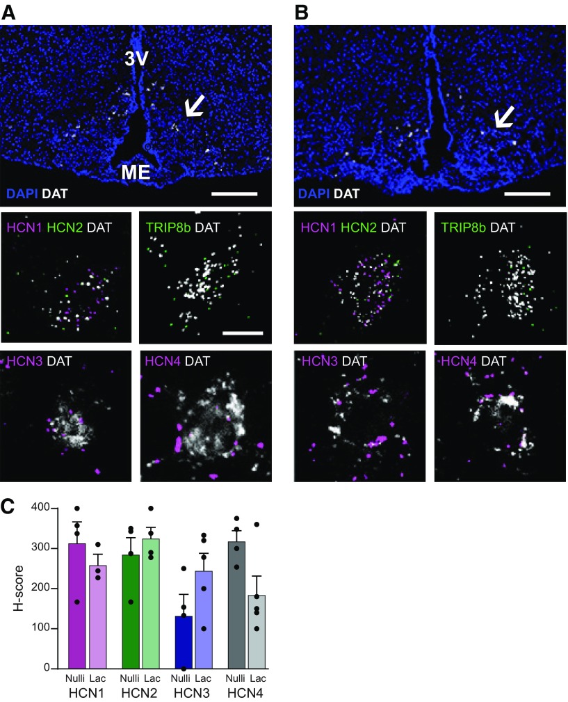
Figure 6.
TIDA neurons express HCN channel mRNA. A, Confocal micrograph from the arcuate nucleus of a female nulliparous mouse in estrus after ISH (RNAScope) performed to detect DAT mRNA (white); section counterstained with DAPI (blue) to visualize anatomy. Note distribution of DAT-expressing TIDA cells in the dmArc (arrow). Insets, High-magnification examples of TIDA cells coexpressing DAT (white) and (in clockwise order): the HCN channel 1 (HCN1; magenta) and HCN2 (green); the HCN channel auxiliary subunit, tetratricopeptide repeat-containing Rab8b-interacting protein (TRIP8b; green); HCN3 (magenta); and HCN4 (magenta). B, Confocal micrograph organized as in A, from the arcuate nucleus of a lactating dam. All RNA transcripts detected in the nulliparous female TIDA neurons can also be found in the same neurons from a lactating female. Scale bars: A, B, 100 μm; Inset, 20 μm. 3V, Third ventricle; ME, median eminence. C, Histogram displaying the H-score (see Materials and Methods) of DAT-expressing neurons in the dmArc that coexpress HCN1-4 mRNA. The individual values represent the score from slices from 3 animals in each group. The H-score is not statistically different for either HCN mRNA between the two experimental groups (unpaired Student's t test: HCN1, p = 0.43; HCN2, p = 0.45; HCN3, p = 0.13; HCN4, p = 0.05).

