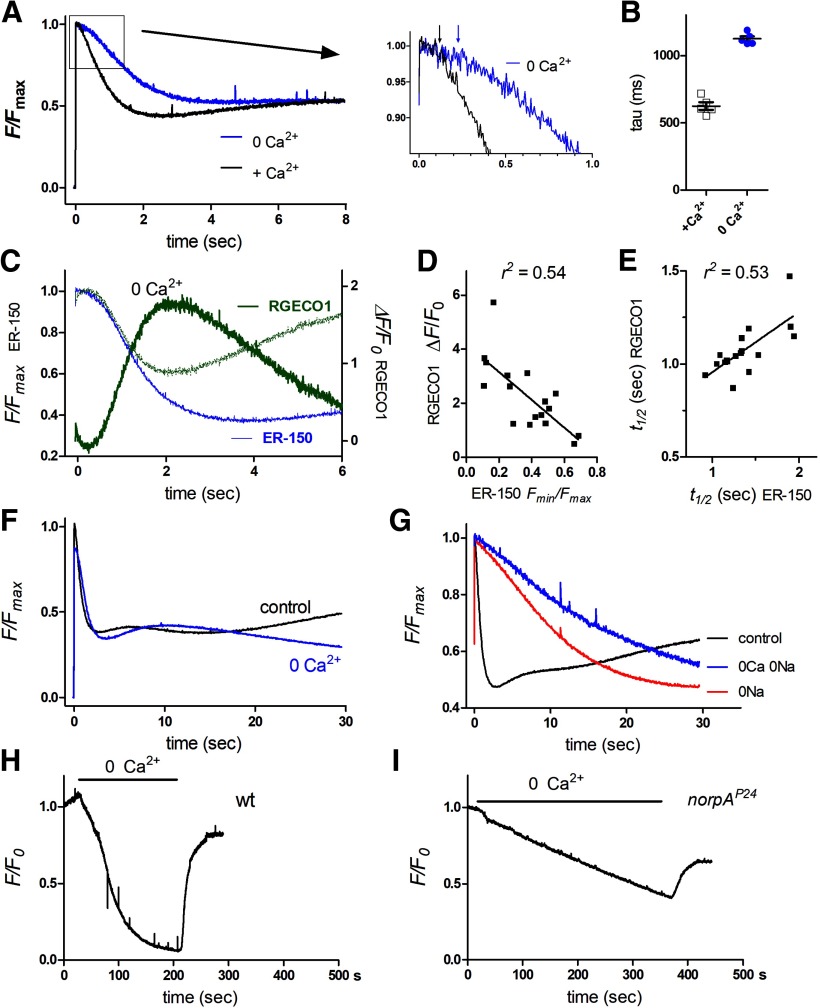
Figure 4.
ER store depletion in dissociated ommatidia in Ca2+-free solutions. A, Normalized ER-150 fluorescence in response to blue excitation control bath and in the same ommatidia perfused for ∼30 s with Ca2+-free bath (0 Ca2+, 1 mm EGTA). Average traces from five ommatidia in three otherwise wild-type flies. Inset (right), Expanded scale to show increase in latency (arrows). B, Time constants (tau) of rapid depletion from exponential fits to decay. C, Store depletion (ER-150 fluorescence, blue trace, expressed as F/Fmax) and cytosolic Ca2+ (RGECO1 fluorescence; green trace expressed as ΔF/F0) measured from the same ommatidia (average of 17 ommatidia from 5 otherwise wild-type flies). Stippled green trace: RGECO1 data replotted after inverting and rescaling to compare time courses. D, Peak RGECO1 ΔF/F0 values (cytosolic Ca2+) from these ommatidia correlated strongly with the extent of store depletion (ER-150 Fmin/Fmax values) in the same ommatidium. E, Time to 50% depletion (t1/2 ER-150) in these ommatidia also correlated with the t1/2 for the rise in cytosolic Ca2+. F, Store depletion (ER-150 fluorescence): averaged from 10 ommatidia from 2 wild-type flies in control bath and during 0 Ca2+ perfusion, monitored over 30 s. G, Store depletion (ER-150 fluorescence) measured from wild-type ommatidia over 30 s in control bath, and same ommatidia during perfusion with 0 Na (130NMDG, 1.5 Ca, 4 Mg; n = 8) and 0Ca/0Na (130NMDG, 0Ca 1 mm EGTA, 4 Mg; n = 3). H, ER-150 fluorescence (normalized to fluorescence at time 0) measured continuously during perfusion with 0 Ca2+ (1 mm EGTA) in a wild-type background; the ER stores depleted almost completely within ∼3 min and then rapidly refilled on re-exposure to normal Ca2+ containing bath. I, During 0 Ca2+ perfusion in a PLC-null (norpAP24) background, store Ca2+ declined more slowly, recovering partially on reperfusion with Ca2+ containing bath.

