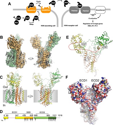
Fig. 1. Structure of the D. melanogaster Dispatched.

(A) Schematic representation of Hh ligand release, which initiates the Hh pathway. (B) A cryo-EM map of Dispatched reconstructed at 3.2-Å resolution; orange and green colors indicate the N- and C-terminal halves of the protein; elements of the density map assigned to bound sterols or unassigned (i.e., corresponding to the unstructured components of the detergent micelle) are colored gray. (C) Atomic model of Dispatched, built using the 3.2-Å map shown in (B). (D) Sequence coverage of the atomic model and annotation of the sequence. The colors of the linear representation of Dispatched sequence in (D) correspond to the colors of the secondary structure elements in (C). Dashed line indicates a portion of the protein not resolved in the cryo-EM map. (E) The view of the protein model from the extracellular side indicates the Cα atoms of the residues E90 and S247; the red dashed line indicates an unresolved loop between these two residues. (F) The space-filling representation of the Dispatched model reveals a large cavity formed by the two ECDs, with the SSD in close proximity.
