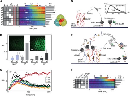
Fig. 4. Late stages of on-chip E. coli SSU assembly.

(A) r-RNA:S2-HA signals for different combinations of SSU domains (a1 to a8), depicted as dynamic color maps. Labels are as in Fig. 3. (B) Averaged fmax values of r-RNA:S2-HA signals for different assembly factor combinations with and without all r-proteins. Error bars represent SD of three repeats. Scale bar, 100 μm. (C) Signal dynamics of r-RNA:S2-HA interactions in the presence of four factors and domain combinations. (D) Scheme: Ribosomes localized on surface antibodies actively engaged in GFP-SecM synthesis with purified SSU added from bulk solution. mRNA is produced from nearby DNA brushes. Inset: TIRF dynamic signal of GFP-secM on surface-bound ribosomes. (E) Scheme: Synthesis and assembly of nascent SSU and binding to surface immobilized LSUs. (F) Signal dynamics color maps of nascent SSU binding onto surface-bound LSU, dependent on different r-protein combinations (b1 to b4). In each panel, the presence of assembly factors is marked as 2F (Era and RsgA), 4F (RbfA, RimM, RimN, and RimP), or 6F (all).
