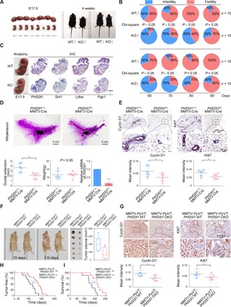
Fig. 6. Phf20l1 deletion induces growth retardation and mammary ductal outgrowth delay and inhibits breast cancer in vivo.

(A) Uterine tissue excised from a pregnant female at 17.5 days post coitum (left). Phf20l1 KO adults are smaller than normal at about 4 weeks old (right). E17.5, embryonic day 17.5. (B) Phf20l1-null mice has delayed reproductive age and exhibited lower fertility. (C) The expression profiles of indicated GRGs were measured using IHC staining in littermate embryos. (D) Mammary ductal developmental defects in Phf20l1 CKO mice (represented as Phf20l1f/f; MMTV-Cre) at about 6 weeks old. (E) IHC staining of cyclin D1 and Ki67 in mammary glands of 6-week-old control and Phf20l1-null mice. (F) Representative bright-field imaging of mammary adenocarcinoma from MMTV-PyVT; Phf20l1f/f; MMTV-Cre (represented as MMTV-PyVT; Phf20l1 CKO) and MMTV-PyVT; Phf20l1+/+; MMTV-Cre (represented as MMTV-PyVT; Phf20l1 WT) mice. The circles indicate surface tumors. The biggest tumors of 110-day-old mice were obtained and calculated (n = 6). (G) IHC staining of Ki67 and cyclin D1 in mammary tumors isolated from MMTV-PyVT; Phf20l1 CKO and MMTV-PyVT; Phf20l1 WT mice. Data shown are means ± SD. Two-tailed unpaired t test, *P < 0.05 and **P < 0.01 (D to G). (H) Mammary adenocarcinoma incidence in MMTV-PyVT; Phf20l1 WT (n = 10) and MMTV-PyVT; Phf20l1 CKO (n = 16) mice depicted as the percentage of tumor-free mice. Mice were considered to be tumor free until a palpable mass (>4.0 mm) persisted for longer than 4 days. Log-rank test was used. (I) Overall survival analysis of the MMTV-PyVT; Phf20l1 WT (n = 9), MMTV-PyVT; Phf20l1 CKO (n = 8) mice, log-rank test. Photo credit: Yongqiang Hou, Tianjin Medical University.
