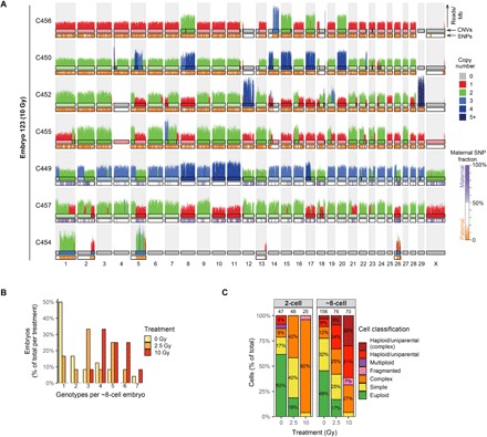
Fig. 4. Chaotic mosaicism in eight-cell–stage embryos produced with damaged sperm.

(A) Standard single-cell WGS derived copy number profiles of seven cells from embryo E123 showing complex genomic abnormalities. Cells C450, C452, C455, and C456 are uniparental with only paternal chromosomes, whereas C449 and C457 are biparental. C454 is a fragmented cell containing chromosomal fragments that are complementary to copy number losses in C449 and C452. The insides of the top chromosome ideograms are color-coded to indicate copy number (see legend for the copy number states). The ideograms for the bottom chromosome ideograms are color-coded to indicate the contribution of maternal SNVs (see legend for details). (B) Embryos produced with damaged sperm frequently show more than three different genetic lineages around the eight-cell stage of development, indicative of genomic instability through mitotic errors. (C) Classification of all the cells for the different treatment groups. The proportion of cells with multiple genomic abnormalities increases with sperm radiation dose. Cells without chromosomal or segmental abnormalities are classified as euploid. Cells with one or two chromosomal or segmental abnormalities are classified as simple. Cells with three or more chromosomal or segmental abnormalities are classified as complex. Fragmented cells only contain a few chromosomal fragments. Numbers above the bars indicate the number of analyzed cells per group.
