
Fig. 3.
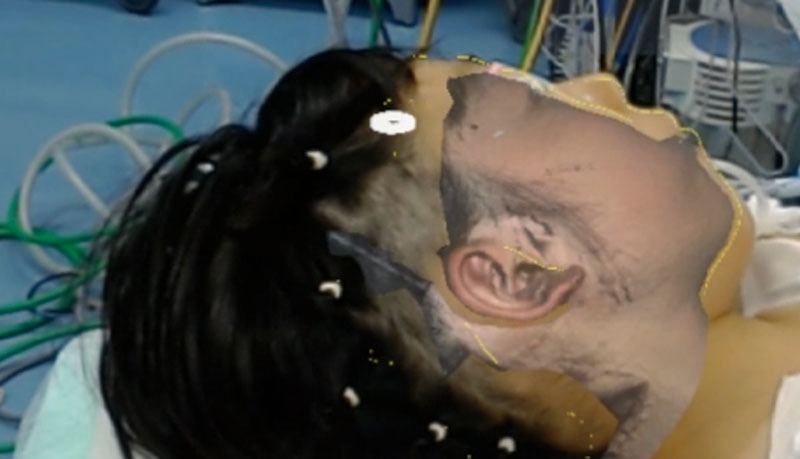
The surgical site seen through HoloLens. The right and left reversed 3D image of the nonaffected auricle was displayed and the outline of the opposite auricle was transferred.
Official websites use .gov
A
.gov website belongs to an official
government organization in the United States.
Secure .gov websites use HTTPS
A lock (
) or https:// means you've safely
connected to the .gov website. Share sensitive
information only on official, secure websites.
The surgical site seen through HoloLens. The right and left reversed 3D image of the nonaffected auricle was displayed and the outline of the opposite auricle was transferred.