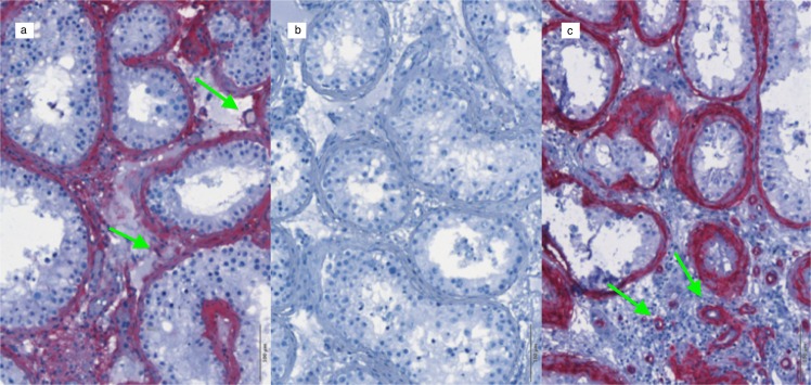
Fig. 2.
Validation protocol for AMHR-2 IHC staining. Tissue sections from human testis; ×20 magnification. a 1:500 AMHR-2 staining and hematoxylin counterstaining; b primary antibody omitted, using hematoxylin counterstaining only; c 1:500 α-SMA staining and hematoxylin counterstaining. The green arrows point towards blood vessels

