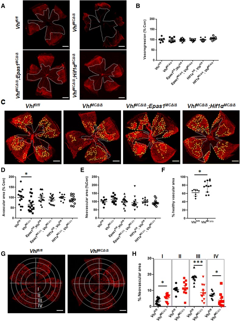
Fig. 1.
Stabilization of HIFs in myeloid cells promotes retinal revascularization in OIR. a Representative pictures of flat-mounted retinas from VhlMCΔ/Δ, VhlMCΔ/ΔEpas1MCΔ/Δ, and VhlMCΔ/ΔHif1aMCΔ/Δ mice at P11 after hyperoxic exposure. b Retinal vasoregression (white depicted area) was similar in all the models after hyperoxic exposure (P11). c Representative pictures of flat-mounted retinas from VhlMCΔ/Δ, VhlMCΔ/ΔEpas1MCΔ/Δ, and VhlMCΔ/ΔHif1aMCΔ/Δ mice at P16 after OIR. d Remaining avascular area was significantly reduced (by 35%) at P16 in retinas from VhlMCΔ/Δ mice but not in the double knock-out mice, although e the total neovascular area (yellow depicted area) remained unchanged in all the models. fVhlMCΔ/Δ retinas showed increased (25%) healthy vascular area at P16 after OIR, when compared with control littermates. g Concentrical division of retinal area revealed h reduced neovascular area (~ 50% reduction) in the peripheral retinas from VhlMCΔ/Δ mice. Scale bars: 0.5 mm. n = 6–14 per group. Data are expressed as mean ± SEM. Values in b, d, and e are represented as % of control for each mouse model. Statistical analysis was performed by one-way ANOVA (b, d, e) and the two-sided Mann–Whitney test (f, h), *p < 0.05

