Fig. 3.
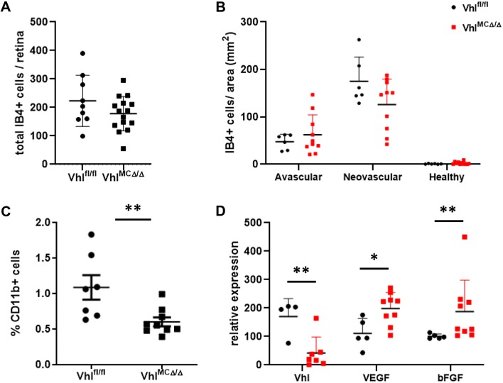
Vhl-deficient myeloid cells produced increased levels of VEGF and bFGF. Quantification of a total lectin-positive myeloid cells per retina and b lectin-positive myeloid cells per area revealed a trend for reduced numbers in neovascular areas of VhlMCΔ/Δ retinas. c Reduced population by 55% of CD11b-positive cells in VhlMCΔ/Δ retinas at P16 after OIR. d CD11b+ cells sorted from VhlMCΔ/Δ retinas showing depletion of Vhl, expressed more VEGF and bFGF (~ 1.8-fold increase) as measured by qPCR at P16 after OIR, when compared with cells sorted from control littermates. n = 4–15 per group. Data are expressed as mean ± SEM. Statistical analysis was performed by two-sided Mann–Whitney test, *p < 0.05, **p < 0.01

