Fig. 2.
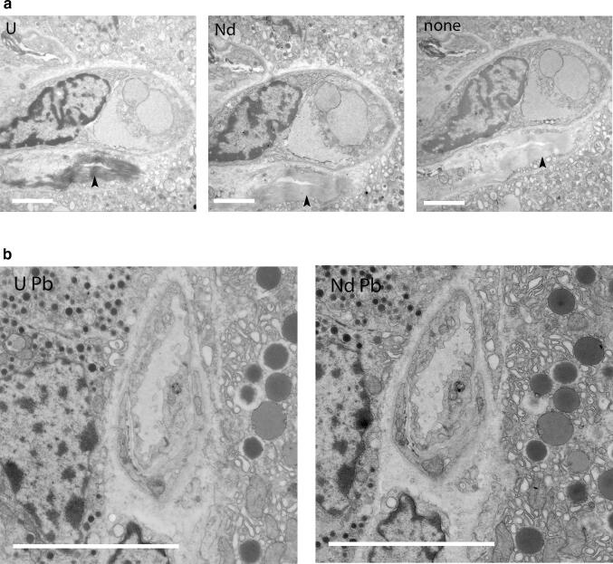
Neodymium Acetate replacing uranyl acetate in durcupan embedded sections, also combined with Reynolds lead citrate. a TEM images of 100 nm sections rat pancreas embedded in Durcupan stained with either 2% uranyl acetate, 4% neodymium acetate or without any stain showing an increased contrast of UAc and NdAc compared to no staining. To visualize the difference in contrast images were recorded with same beam spreading and fixed scaling. Note the equal contrast for UAc and NdAc, only collagen being more dark with UAc (arrow heads). Bars: 2 µm. b TEM images of 100 nm sections rat pancreas embedded in Durcupan stained with either 2% uranyl acetate, 4% neodymium acetate followed by Reynolds Lead Citrate contrasting give very similar contrast. Bars: 5 µm

