Figure 5.
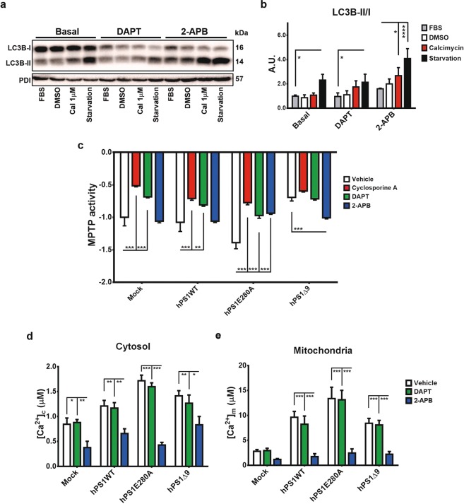
γ- secretase dependent and independent cellular stress response in hPS1E280A cells. (a) LC3B conjugation was evaluated in hPS1E280A cells treated with γ-secretase inhibitor DAPT and ER calcium channels inhibitor 2-APB. Representative western blot for LC3B from hPS1E280A N2a cells treated with FBS, DMSO, calcimycin and serum starved, all for 16 h. Cells were assessed with and without concurrent 16 h exposure to DAPT or 2-APB. (b) Bar graphs for densitometric analysis of hPS1E280A N2a cells. DAPT increased LC3B conjugation when compared to basal conditions and 2ABP increased it after calcymicin treatment or starvation in hPS1E280A cells. (c) Mock transfected, hPS1WT, hPS1E280A, and hPS1Δ9 N2a cells were challenged with 1 μM ionomycin to induce MPTP opening and quenching of the calcein signal. Cells were treated with Cyclosporin A, DAPT, or 2-APB. Cyclosporin A and DAPT inhibited MPTP opening in mock, hPS1WT, and hPS1E280A cells while 2-APB only showed an effect in PS1 mutants, inhibiting MPTP opening in hPS1E280A cells and accelerating it in hPS1Δ9 cells. (d) Bar graphs of maximum cytosolic calcium concentration and (e) mitochondrial calcium concentration in the different N2a cell lines, treated with DMSO (vehicle), DAPT and 2-APB for 16 h. 2-APB decreased mitochondrial calcium levels in PS1 overexpressing cells and cytoplasmic calcium levels in all cells. *P < 0.05, **P < 0.01, ***P < 0.001. Data are mean ± SEM, Two-Way ANOVA.

