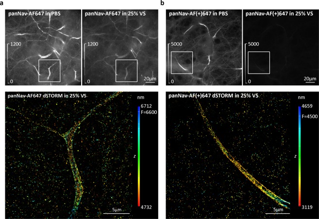
Figure 6.
Comparison of the effect of 25% Vectashield (25% VS) on AF647 and AF(+)647 fluorescence. (a) Mouse cortical neurons (MCN) immunolabelled with anti-pan voltage-gated sodium channels (panNav) antibody, followed by AF647-conjugated secondary antibody. (b) MCN immunolabelled with panNav, followed by AF(+)647-conjugated secondary antibody. Upper panels show widefield images of the same field of view acquired in PBS and in 25% VS. Brightness and contrast were linearly adjusted to show the same display range in both PBS and 25% VS conditions (as indicated by look-up table (LUT) intensity scale bars). Lower panels show corresponding 3D dSTORM images of the boxed regions from panels (a and b). The z positions in the 3D dSTORM images are colour-coded according to the height maps shown on right. Height maps contain minimal, maximal and focal (F) z position values.

