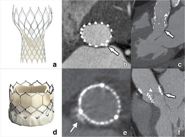
Fig. 1.
CT images of a Medtronic self-expandable Corevalve (a) and Edwards Lifesciences balloon-expandable Sapien valve (d). After deployment, a self-expandable valve will conform itself to the normal annular contour, acquiring an oval cross-sectional morphology (b). Compared with a balloon-expandable valve, the self-expandable Corevalve is larger in size and contains an inflow (I), waist (w), and outflow (O) functional part (c). This outflow part is by design intended to extend into the ascending aorta, covering but not obstructing the coronary ostia. The balloon-expandable Sapien valve (d) is shorter, with a mostly circular cross-sectional contour after deployment (e) as it forces this circular morphology on the annulus through the radial forces of the expanding balloon. In contrast to the self-expandable Corevalve, it remains within the aortic sinus (f). Within both THVs, the pericardial leaflets can be appreciated as fine hypodense linear structures (arrowheads in c, f), its visibility dependent on image quality. Small interposing calcifications (arrows in b, e) have in these examples only a minimal effect on THV expansion. Reused from reference [40], with permission

