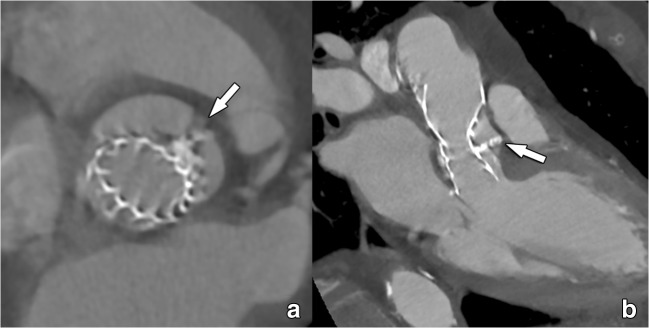
Fig. 5.
Incomplete and asymmetric deployment of a self-expandable THV due to interposition of extensive native leaflet calcifications (arrow in a, b) between the prosthetic valve and the wall of the aortic sinus. Severe calcifications can complicate prosthesis deployment as in this case, leading to a deformed THV. Nevertheless, caution should be taken when extrapolating morphological findings into a potential dysfunction. While in this case the residual gap between the THV and the aortic wall would suggest a severe paravalvular leakage, this was not the case on Doppler echocardiography examination, with the calcification apparently acting as an additional seal. Valvular function was acceptable, and no further intervention was deemed necessary

