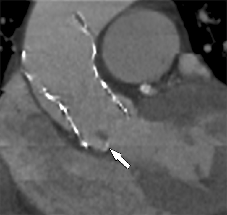
Fig. 6.

Incorrect positioned THV, which is tilted and does not fully extend into the aortic annulus. As such, parts of the native right aortic valve leaflet is protruding into the inflow part of this self-expandable THV (arrow), causing a residual valve gradient on Doppler echocardiography. CT is very useful in detecting the cause of THV dysfunction in cases where Doppler echocardiography does not provide an answer. In this case, function was improved after balloon dilatation of the inflow part of the THV, further crushing the remaining valve leaflets against the adjacent aortic wall
