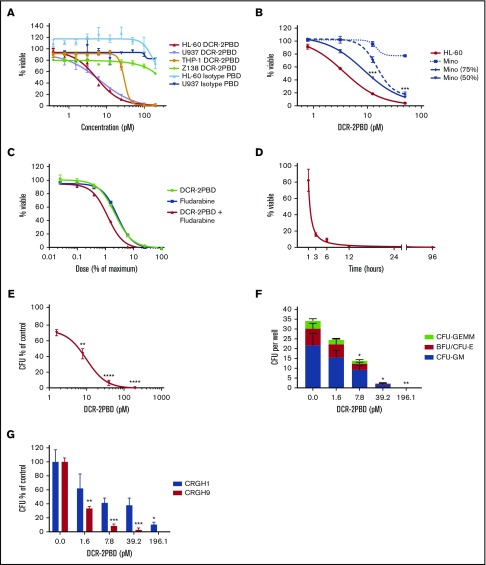
Figure 3.
DCR-2-PBD in vitro cytotoxicity. (A) Inhibitory concentration curves on CD300f+ AML cell lines (HL-60, U937, and THP-1) and CD300f− lymphoma cell line (Z-138) using DCR-2-PBD or isotype-PBD (performed in triplicate). (B) Bystander killing assay using an CD300f− lymphoma cell line (Mino) and HL-60 either alone or in combination. In combination conditions, only the percentage of viable of Mino cells is shown (performed in triplicate) (***P < .001 Mino 50% at 25 and 12.5 pM, Mino 75% at 25 pM). (C) Combination DCR-2-PBD and fludarabine inhibitory concentration curves using HL-60 (performed in triplicate). (D) Time-dependent killing assay of DCR-2-PBD on HL-60, with final viability measured at 96 hours. (E) Total CFU inhibitor concentration curve with DCR-2-PBD (3 CB samples each performed in duplicate) (**P < .01 CFU inhibition at 7.8 pM, ****P < .0001 CFU inhibition at 39.2 and 196.1 pM). (F) Individual CFU subtype formation inhibition by DCR-2-PBD (*P < .05 CFU inhibition at 7.8 and 39.2 pM, **P < .01 CFU inhibition at 196.1 pM). (G) Individual AML CFU inhibition with DCR-2 PBD (performed in duplicate) (*P < .05 CFU inhibition at 196.2 pM for CRGH1, **P < .01 CFU inhibition at 1.6 pM for CRGH9, ***P < .001 CFU inhibition at 39.2 pM and 196.2 pM for CRGH9). BFU, burst-forming unit; GEMM, granulocyte, erythroid, macrophage, megakaryocyte; GM, granulocyte macrophage.

