Figure 5.
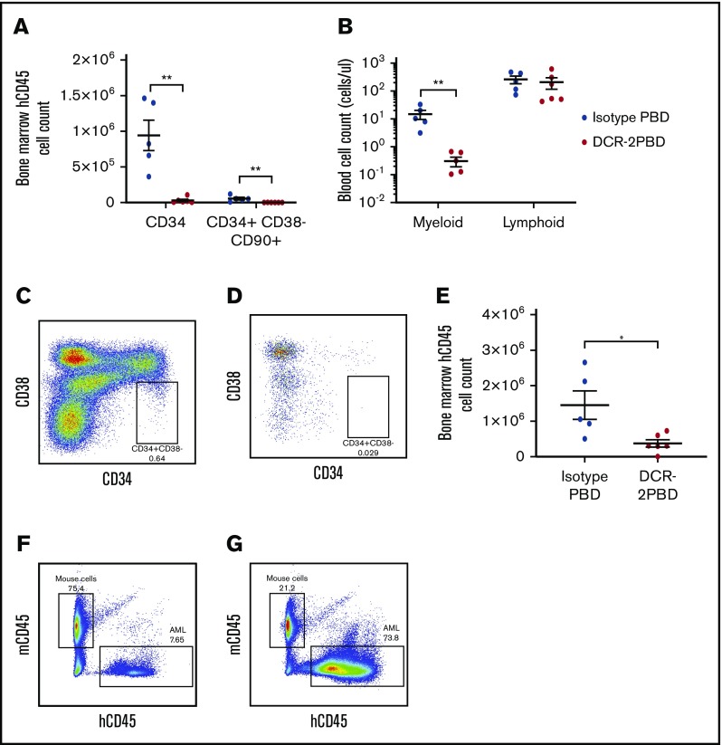
DCR-2-PBD depletes HSPCs in vivo. (A) Total cell count of CD34+ and CD34+ CD38−CD90+ cells in humanized NSG mouse BM 7 days after injection of DCR-2-PBD (n = 6) or isotype-PBD (n = 5) (**P < .01 reduction in CD34+ and CD34+ CD38−CD90+ cells in the DCR-2-PBD cohort compared with the isotype control cohort). (B) Cell count per microliter of blood from DCR-2-PBD and isotype-PBD treated humanized mice (**P < .01 reduction in myeloid cells in PB of DCR-2-PBD cohort compared with isotype-PBD cohort). (C-D) Representative plots of human CD45+ cells in the BM of a control humanized mouse (C) and a DCR-2-PBD treated humanized mouse (D). (E) BM enumeration of primary AML 6 days after injection of mice treated with isotype-PBD and DCR-2-PBD (n = 6 DCR-2-PBD, n = 5 isotype-PBD) (*P < .05 reduction of primary AML cells in BM, DCR-2-PBD cohort compared with the isotype-PBD cohort). (F-G) Representative plots of mouse CD45 vs human CD45 cells in mice engrafted with primary AML treated with isotype-PBD (F) and DCR-2-PBD (G).

