
Figure 2.
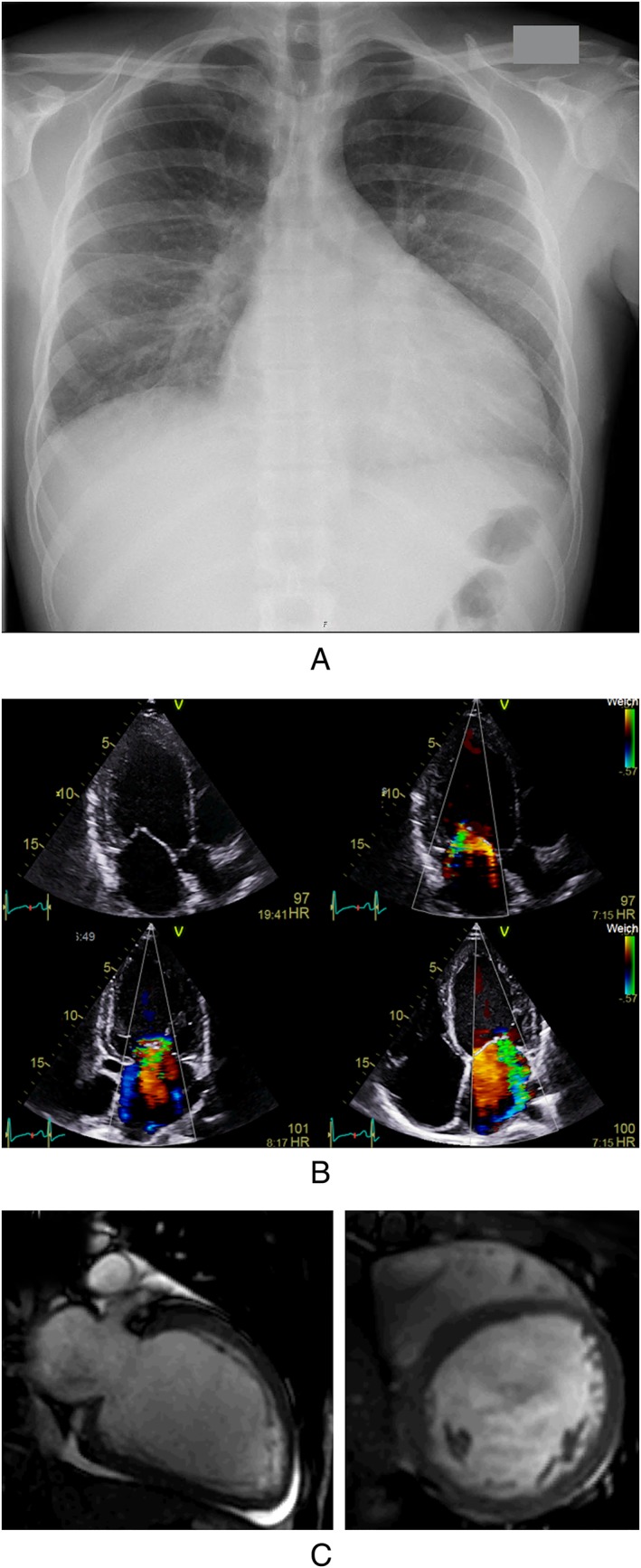
Clinical case report of a patient with frequent MA abuse. A: Chest X‐ray showing cardiomegaly and lung congestion at the time of hospital admission. B: Transthoracic echocardiogram showing LV‐dilatation and moderate to severe secondary mitral regurgitation due to ring dilatation. C: Diastolic 2 chamber and shortaxis views in cardiac MRI showing LV enlargement without signs of myocardial infarction or inflammation and discrete pericardial effusion.