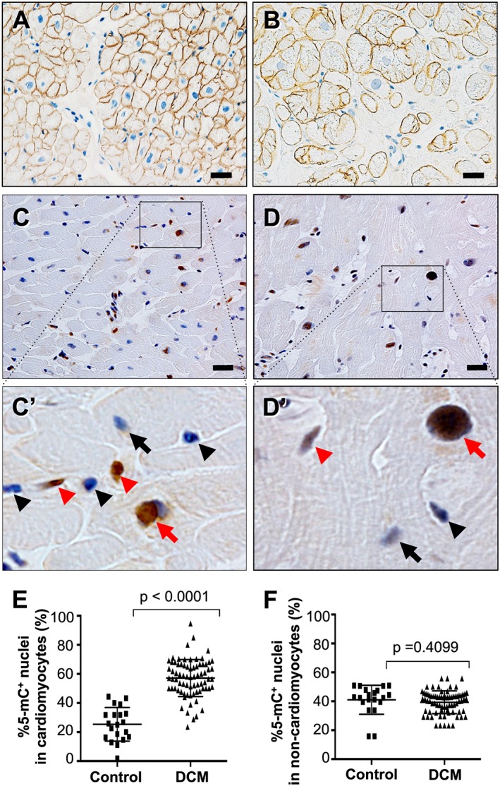
Figure 1.

Cell type specification and immunohistochemical detection of 5‐mC in endomyocardial biopsy specimens at the light microscopic level. (A and B) Immunohistochemistry for dystrophin in a control specimen in panel A and a DCM specimen in panel B. Plasma membranes and T‐tubules are stained specifically in cardiomyocytes. Scale bars, 20 μm. (C and D) 5‐mC‐positive nuclei are stained brown while 5‐mC‐negative nuclei are stained blue in both a control specimen in panel C and a DCM specimen in panel D. Panels A′ and B′ are highly magnified images of the boxed areas in panels A and B, respectively. Red arrows, 5‐mC‐positive cardiomyocyte nuclei; black arrows, 5‐mC‐negative cardiomyocyte nuclei; red arrowheads, 5‐mC‐positive non‐cardiomyocyte nuclei; black arrowheads, 5‐mC‐negative non‐cardiomyocyte nuclei. Scale bars, 20 μm. (E) Graph showing comparison of the %5‐mC‐positive nuclei in cardiomyocytes between controls (n = 20) and DCM patients (n = 75). (F) Graph showing comparison of the %5‐mC‐positive nuclei in non‐cardiomyocyte interstitial cells between controls (n = 20) and DCM patients (n = 75).
