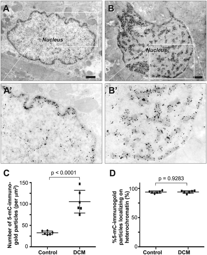
Figure 2.

Immunocytochemical detection of 5‐mC in cardiomyocytes in endomyocardial biopsy specimens at the electron microscopic level. Immunogold particles bound to 5‐mC localize predominantly in heterochromatin in both normal (A) and bizarrely shaped (B) nuclei. The bizarrely shaped nucleus is rich in heterochromatin where immunogold particles are abundant. Panels A′ and B′ are highly magnified and lightly printed images of the boxed areas in panels A and B, respectively. They highlight the relationship between heterochromatin and immunogold particles. Scale bars, 1 μm. (C) Graph showing comparison of the number of 5‐mC bound immunogold particles between controls (n = 6) and DCM patients (n = 6). (D) Graph showing fraction of the 5‐mC bound immunogold particles localized on heterochromatin in controls (n = 6) and DCM patients (n = 6).
