Figure 3.

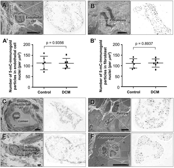
Immunocytochemical detection of 5‐mC in non‐cardiomyocyte interstitial cells in endomyocardial biopsy specimens from patients with DCM. (A) Capillary endothelial cell. (B) Fibroblast. (A′ and B′) Graphs showing comparison of the number of 5‐mC bound immunogold particles in capillary endothelial cell nuclei (A′) and in fibroblast nuclei (B′) between controls (n = 6) and DCM patients (n = 6). Group comparisons were made using Student's t‐tests. (C) Smooth muscle cell in a small artery. (D) Pericyte in an arteriole. (E) Macrophage. (F) Polymorphonuclear cell within a vascular lumen. Scale bars, 1 μm.
