Figure 4.
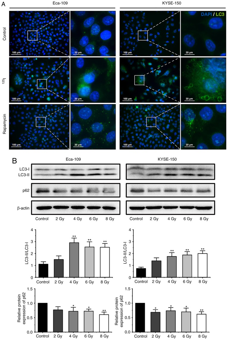
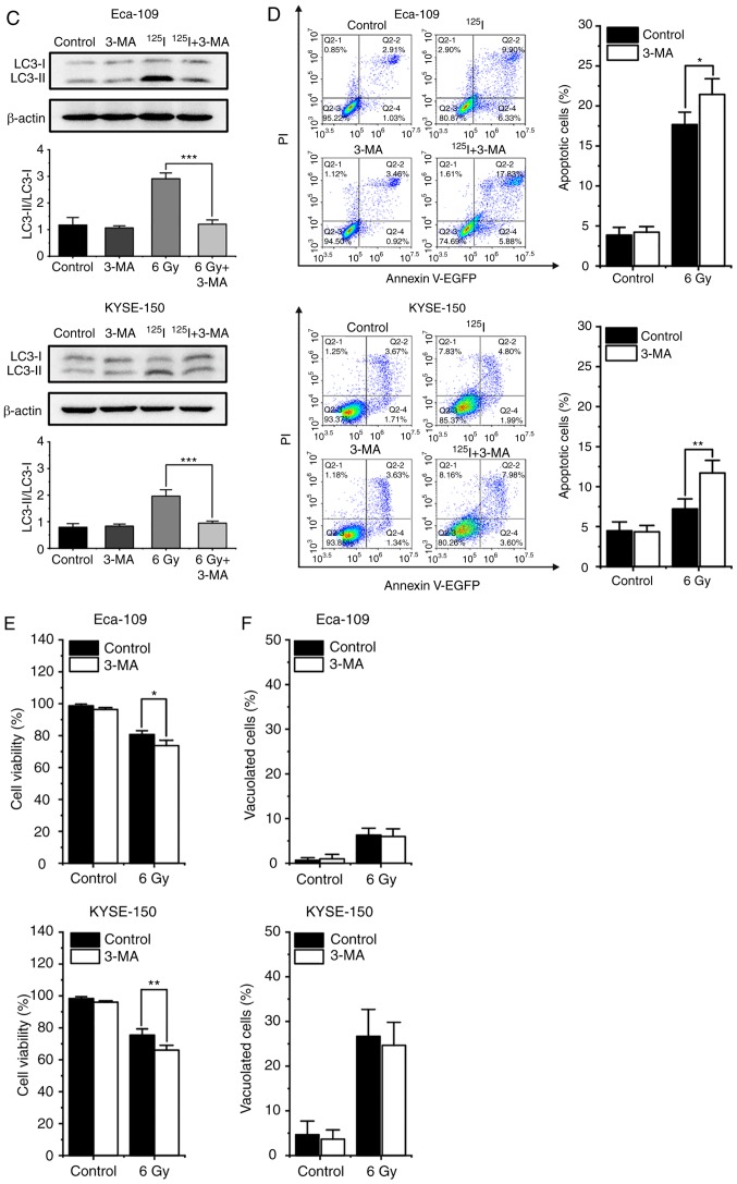
125I seed radiation induces protective autophagy in Eca-109 and KYSE-150 cells. (A) Following 6 Gy irradiation, and 48 h of culture, LC3 punctuation (green) was detected using a fluorescent microscope. Nuclei were stained with DAPI (blue). Cells treated with 200 nM rapamycin overnight were used as the positive control. (B) Cells were exposed to 2, 4, 6 or 8 Gy radiation. The ratios of LC3-II to LC3-I expression, and the relative protein expression of p62 were analyzed by western blot. β-actin was used as a loading control. Cells were pretreated with or without 3-MA, 2 h prior to 6 Gy irradiation. After 48 h of the indicated treatment, (C) the ratios of LC3-II to LC3-I were determined by western blot, and (D) apoptosis was analyzed by Annexin V-EGFP/PI assay using flow cytometry. (E) The percentage of cells with trypan blue exclusion, and (F) the percentage of vacuolated cells were measured under a light microscope. *P<0.05, **P<0.01, ***P<0.001 vs. control group. 125I, Iodine-125; 3-MA, 3-methyladenine; EGFP, enhanced green fluorescent protein; PI, propidium iodide.


